
See it in 3D. Each chapter has a practice quiz and study tips for learning the topic.
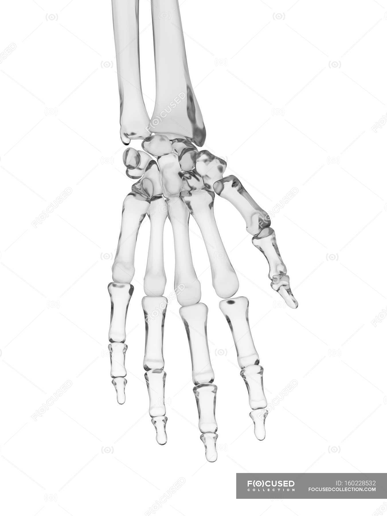
Human Hand Bones Structure Fingers Artwork Stock Photo 160228532
Hand Wikipedia

Metacarpal Bones Wikipedia
It is the branch of science that deals with the study of the skeletal system their structure and functions.
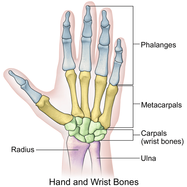
Hand skeletal structure. Thorax is a Plan S compliant Transformative Journal. This article the second in a two-part series considers the structure and function of the musculoskeletal system reviews the structure of muscles and joints and identifies some of the common pathology occurring at these structures. Muscle organ and skeletal anatomy.
What would happen if humans did not have bones. The scaphoid is at particular risk of avascular necrosis after fracture because of its so-called. About 200-250 muscle fibers are surrounded by endomysium forming the functional unit of the muscle the primary bundle.
How do bones move. Most of the cell is occupied by striated thread-like myofibrils. A sarcomere or muscle functional unit extends from Z line to Z line.
What are bones made up. The skeletal system supports and protects the body while giving it shape and form. U pper Extrem eties shoulder pectoral girdle upper and lower arm w rist and hand 1.
The cavities or spaces of the body contain the internal organs or visceraThe two main cavities are called the ventral and dorsal cavities. Koehlerzimmers borderlands of normal and early pathological findings in skeletal radiography. In the specific case of striated muscles contraction and relaxation mechanisms are both regulated by rapid changes in myoplasmic free Ca 2.
Free multiple-choice quizzes on the skeletal system of the human body including the bones of the full and axial skeletons the bones of the hand and foot and the anatomy of bones. Skeletal musculature Structure of the skeletal muscle. What are skeletal formulae or skeletal structures of organic compounds.
Any part of a body that engages in voluntary movements such as your hand the wings of a bird the trunk of an elephant all require the use of skeletal muscles. Many mammals and other animals have grasping appendages similar in form to a hand such as paws claws and talons but these are not scientifically considered to be grasping handsThe scientific use of the term hand in this sense to distinguish the terminations of the front paws from the hind ones is an example of anthropomorphism. Just call me Blob.
Dislocations fractures herniated disc infectious arthritis osteoarthritis osteoporosis. Muscle fibers and connective tissue layers make up the skeletal muscleA skeletal muscle fiber is around 20-100 µm thick and up to 20 cm longEmbryologically. They have a radius and ulna as well as a complete hand structure including five phalanges or finger bones.
The scaphoid bone of the hand is the most commonly fractured carpal bone typically by falling on an outstretched hand FOOSH. Clarification needed The term comes from Greek σκελετός skeletós. Plus there are links to lots of other great anatomy and physiology quizzes and other resources.
It develops by the chain-like fusion of myoblasts. Structure of Skeletal Muscle. There are several different skeletal types.
The ventral is the larger cavity and is subdivided into two parts thoracic and abdominopelvic cavities by the diaphragm a. Muscle fibers are multinucleated with the nuclei located just under the plasma membrane. Skeletal System Ppt 1.
Well go over the function and anatomy of the skeletal system before diving into. It contains textbook resources such as chapter review guides homework sets tutorials and printable images. In a fracture of the scaphoid the characteristic clinical feature is pain and tenderness in the anatomical snuffbox.
Anatomy of a Dog. Skeletal System Functions Structure - 2 main parts. IBRA the International Bone Research Association for surgery research plastic surgery oral surgery and oral maxillofacial surgery.
The detailing of these structures changes based on dog breed due to the huge variation of size in dog breeds. Within each myofibril there are dense Z lines. The capacity is less.
They look very bare because in skeletal formulae the hydrogen atoms attached directly to carbons are removed leaving just a carbon skeleton with functional groups attached to it. This site was designed for students of anatomy and physiology. Dog anatomy details the various structures of canines eg.
Walker J 2020 Skeletal system 2. You will describe the structure and function of the skeletal system. How many bones do humans have.
The bones of the human skeletal system are divided into an axial region and an appendicular region. The thoracic cage gives your upper torso structure. Excitable tissue responds to stimuli through electrical signals.
Human Anatomy Physiology. SKELETAL SYSTEM COMPOSED OF. The skeletal system is the foundation of your body giving it structure and allowing for movement.
Visit Kenhub for more skeletal system quizzes. Axial Appendicular Basic Types - 4 kinds Cartilage Joints Ligaments Tendons Functions of the Skeletal System Provides a strong steady frame for the bodys muscle to move. Thorax is one of the worlds leading respiratory medicine journals publishing clinical and experimental research articles on respiratory medicine paediatrics immunology pharmacology pathology and surgeryThorax seeks to publish significant advances in scientific understanding which are likely to impact on clinical practice.
The excitationcontraction coupling ECC mechanism in skeletal muscle. Structure and function of the musculoskeletal system. Each sarcomere has thick and thin filaments.
Inside their pectoral fins dolphins have a skeletal structure similar to a human arm and hand. Women have smaller cages than men. -Bones -Cartilage -Joints -Ligaments 4.
As well as discoloration of the hands weakness of the hand or arm and stiffness. Skeletal muscle is an excitable contractile tissue responsible for maintaining posture and moving the orbits together with the appendicular and axial skeletonsIt attaches to bones and the orbits through tendons. Ca 2 cell homeostasis and signalling result from dynamic interactions between mechanisms that provoke an increase of cytoplasmic free Ca 2 and those that reduce it.
Contractile tissue is able to generate tension of force. Quizzes on human skeletal system anatomy bone anatomy and bone markings. Scapula clavicle only attached to trunk by 1 joint betw een sternum and clavicle scapula is very m oveable Ð acts as alm ost a 4th.
The skeleton is the framework that provides structure to the rest of the body and facilitates movement. In organic chemistry skeletal formulae are the most abbreviated diagrammatic descriptions of molecules in common use. They have a humerus complete with a ball and socket joint.
A skeleton is a structural frame that supports an animal body. The exoskeleton which is the stable outer shell of an organism the endoskeleton which forms the support structure inside the body and the hydroskeleton a flexible skeleton supported by fluid pressure. Ziser Lecture Notes 20104 15 Appendicular Skeleton A.
Human Skeletal System Human Body Facts Skeleton Bones Facts
![]()
Human Hand Bones And Joints Skeleton Vector Sketch Body Anatomy Icon Isolated Symbol Of Arm Wrist And Fingers Limbs Structure Of Shoulder For Anatomi Stock Vector Image Art Alamy
Fig Bones Of A Human Hand And Wrist Adapted From The Human Anatomy Download Scientific Diagram

Bones Of The Human Hand My Poor Right 3rd Distal Phalange Lol Human Anatomy And Physiology Anatomy Anatomy And Physiology
2 350 Hand Bone Stock Photos Pictures Royalty Free Images Istock


Hand Skeleton Structure 5 Download Scientific Diagram
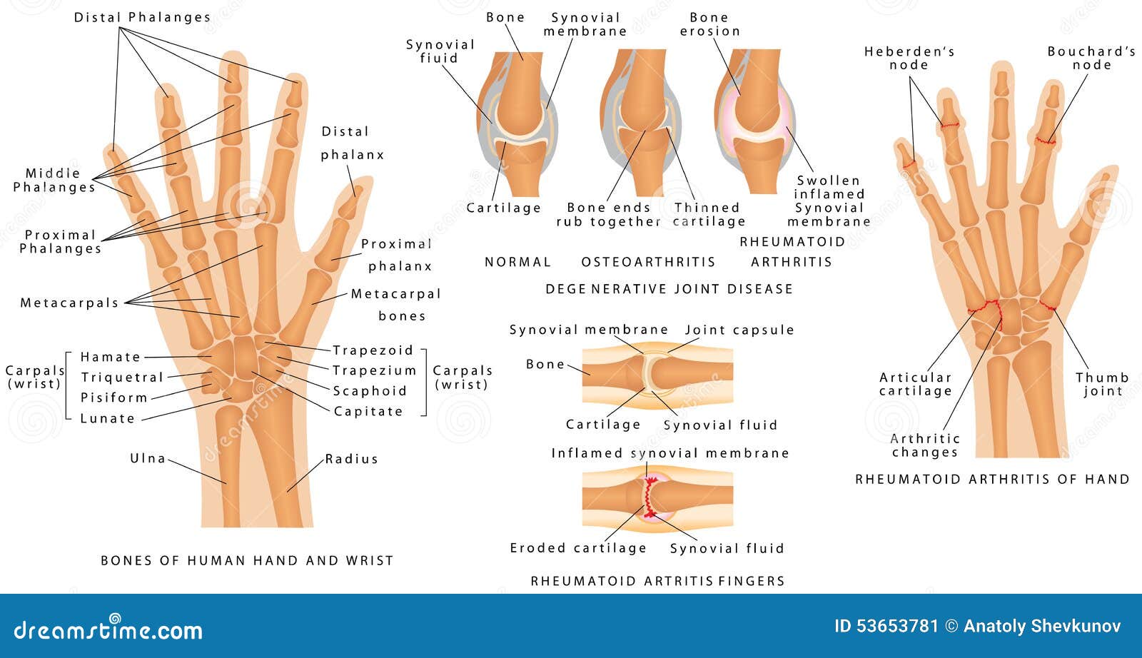
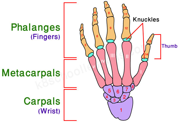
Skeletal System Phalanges Stock Vector Illustration Of Bone 53653781
Hand Anatomy And Function Bone And Spine


